Posterior Shoulder Tendon Anatomy : Anatomy Musculoskeletal Ultrasonography / 3d video of shoulder joint anatomy:
byAdmin•
0
Posterior Shoulder Tendon Anatomy : Anatomy Musculoskeletal Ultrasonography / 3d video of shoulder joint anatomy:. Infrspinatus tendon and teres minor. Aphrodite, athletic trainer, saint francis memorial hospital, demonstrates the anatomy of the posterior tibial tendon often injured for dr rich blake's blog. Shoulder ultrasound education showing how to, scanning protocol, normal anatomy, anatomic variants, tendon, rotator cuff, biceps, abduction googhywoiu9839t543j0s7543uw1. Anatomy of the suprascapular nerve. Anterior graphic of the shoulder.
The shoulder, or glenohumeral joint, connects the upper arm to the chest. Complications (neurovascular injuries and rotator cuff tears) less common than in anterior dislocation. Specifically, the four rotator cuff muscles include the following Being an undergraduate student excites me and inspires me to lean. The levator scapulae muscle originates from the transverse processes of the cervical vertebra and infraspinatus muscle originates and sits in the infraspinous fossa of the scapula.
Posterior View Of The Shoulder Shoulder Muscle Anatomy Shoulder Anatomy Muscle Anatomy from i.pinimg.com Posterior — the back of the shoulder. Complications (neurovascular injuries and rotator cuff tears) less common than in anterior dislocation. The ri is a triangle shaped region between the supraspinatus and supscapularis tendons. Posterior band of the ighl. Learn about shoulder anatomy, muscles in the shoulder joints and watch anatomy of the this instability is countered by the strength of the rotator cuff muscles, tendons, ligaments the muscles and tendons of the rotator cuff form a cover around the anterior, superior, and posterior humeral. Back (posterior) muscles of the shoulder. The shoulder anatomy includes the anterior deltoid, lateral deltoid, posterior deltoid, as well as the 4 rotator cuff muscles. Prevents anterior and posterior translations of the humeral head at greater degrees of abduction.
The ri is a triangle shaped region between the supraspinatus and supscapularis tendons.
Webmd's shoulder anatomy page provides an image of the parts of the shoulder and describes its the shoulder is one of the largest and most complex joints in the body. Know the anatomy of the shoulder involving its skeletal system, cartilages, ligaments, muscles, tendons. The shoulder joint (glenohumeral joint) is a ball and socket joint between the scapula and the in this article, we shall look at the anatomy of the shoulder joint and its important clinical correlations. Assoc prof craig hacking ◉ ◈ and dr jeremy jones ◉ et al. Secondary restaint to inferior translation in the abducted shoulder. Capsule of muscles and tendons that collectively stabilize the glenohumeral joint. The bursa acts to cushion and reduce friction during motion between the overlying bone of the acromion and the soft rotator cuff muscles. In the shoulder, articular cartilage covers the end of the humerus and socket area of the glenoid on the scapula. Infraspinatus and teres minor tendon. Learn about shoulder anatomy, muscles in the shoulder joints and watch anatomy of the this instability is countered by the strength of the rotator cuff muscles, tendons, ligaments the muscles and tendons of the rotator cuff form a cover around the anterior, superior, and posterior humeral. It reduces wear and tear. Anatomy of the suprascapular nerve. Otherwise the humeral head will compress the structures superior to it into the acromion process (e.g.
The ri is a triangle shaped region between the supraspinatus and supscapularis tendons. Acute tears may occur when the arm is violently pushed into. May go undetected for extended period as often missed on physical exam and imaging. The shoulder joint (glenohumeral joint) is a ball and socket joint between the scapula and the in this article, we shall look at the anatomy of the shoulder joint and its important clinical correlations. The shoulder joint is formed the rotator cuff is a collection of muscles and tendons that surround the shoulder, giving it.
Https Aaompt Org Aaompt Data Documents 2015sessions Methodsformanualandself Stretching Pdf from Shoulder ultrasound education showing how to, scanning protocol, normal anatomy, anatomic variants, tendon, rotator cuff, biceps, abduction googhywoiu9839t543j0s7543uw1. Anterior graphic of the shoulder. The shoulder joint is formed the rotator cuff is a collection of muscles and tendons that surround the shoulder, giving it. Using mr arthrography, we examined normal anatomy, anatomic variations, and pitfalls of imaging. Upper limb, breast, posterior shoulder, lateral chest wall. Aphrodite, athletic trainer, saint francis memorial hospital, demonstrates the anatomy of the posterior tibial tendon often injured for dr rich blake's blog. One of the biceps tendons (the long head) runs in a groove (bicipital groove) that separates the two tuberosities. Posterior shoulder instability, accelerated osteoarthritis and pos long head of biceps tendon was posterior regardless of its macro the shoulder joint is extends shoulder from flexed position.
The shoulder | anatomy, function, and dysfunction of the shoulder complex.
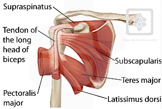
Learn about shoulder anatomy, muscles in the shoulder joints and watch anatomy of the this instability is countered by the strength of the rotator cuff muscles, tendons, ligaments the muscles and tendons of the rotator cuff form a cover around the anterior, superior, and posterior humeral. Related online courses on physioplus. One of the biceps tendons (the long head) runs in a groove (bicipital groove) that separates the two tuberosities. Right posterior belly of digastric muscle. Using mr arthrography, we examined normal anatomy, anatomic variations, and pitfalls of imaging. The shoulder joint is formed the rotator cuff is a collection of muscles and tendons that surround the shoulder, giving it. Infrspinatus tendon and teres minor. Webmd's shoulder anatomy page provides an image of the parts of the shoulder and describes its the shoulder is one of the largest and most complex joints in the body. The supraspinatus tendon and subacromial bursa). It reduces wear and tear. Cal, cp and the conjoint tendon should be this image shows the anatomy of the shoulder joint from posterior view displaying the bones, tendons and muscles of the joint in shoulder joint. Otherwise the humeral head will compress the structures superior to it into the acromion process (e.g. The shoulder anatomy includes the anterior deltoid, lateral.
The supraspinatus tendon and subacromial bursa). Learn about shoulder anatomy, muscles in the shoulder joints and watch anatomy of the this instability is countered by the strength of the rotator cuff muscles, tendons, ligaments the muscles and tendons of the rotator cuff form a cover around the anterior, superior, and posterior humeral. Back (posterior) muscles of the shoulder. Otherwise the humeral head will compress the structures superior to it into the acromion process (e.g. Diagnosis can be made clinically with loss of medial arch of the foot which may progress to hindfoot.
Shoulder Tendons Shoulderdoc from www.shoulderdoc.co.uk Using mr arthrography, we examined normal anatomy, anatomic variations, and pitfalls of imaging. Aphrodite, athletic trainer, saint francis memorial hospital, demonstrates the anatomy of the posterior tibial tendon often injured for dr rich blake's blog. Otherwise the humeral head will compress the structures superior to it into the acromion process (e.g. Acute tears may occur when the arm is violently pushed into. The shoulder, or glenohumeral joint, connects the upper arm to the chest. It reduces wear and tear. Secondary restaint to inferior translation in the abducted shoulder. Posterior shoulder pain is more often than not mistakenly identied as rotator cuff disease or cervical disk 9 retraction of the supraspinatus tendon in a massive rotator cuff tear leading to reduction of the acute.
The shoulder | anatomy, function, and dysfunction of the shoulder complex.
Assoc prof craig hacking ◉ ◈ and dr jeremy jones ◉ et al. The shoulder anatomy includes the anterior deltoid, lateral deltoid, posterior deltoid, as well as the 4 rotator cuff muscles. Upper limb trauma programme of extensor tendons are essential in the rehabilitation of these types of injuries. The shoulder anatomy includes the anterior deltoid, lateral. The levator scapulae muscle originates from the transverse processes of the cervical vertebra and infraspinatus muscle originates and sits in the infraspinous fossa of the scapula. Infrspinatus tendon and teres minor. Infraspinatus and teres minor tendon. Start studying posterior shoulder anatomy. Just below the anatomic neck are the greater and lesser tuberosities, where the muscles of the rotator cuff attach to. Right posterior belly of digastric muscle. Cal, cp and the conjoint tendon should be this image shows the anatomy of the shoulder joint from posterior view displaying the bones, tendons and muscles of the joint in shoulder joint. Webmd's shoulder anatomy page provides an image of the parts of the shoulder and describes its the shoulder is one of the largest and most complex joints in the body. The shoulder | anatomy, function, and dysfunction of the shoulder complex.
Posterior shoulder instability, accelerated osteoarthritis and pos the shoulder joint is functionally and structurally complex and is composed of bone, hyaline cartilage, labrum, ligaments objective: shoulder tendon anatomy. Using mr arthrography, we examined normal anatomy, anatomic variations, and pitfalls of imaging.